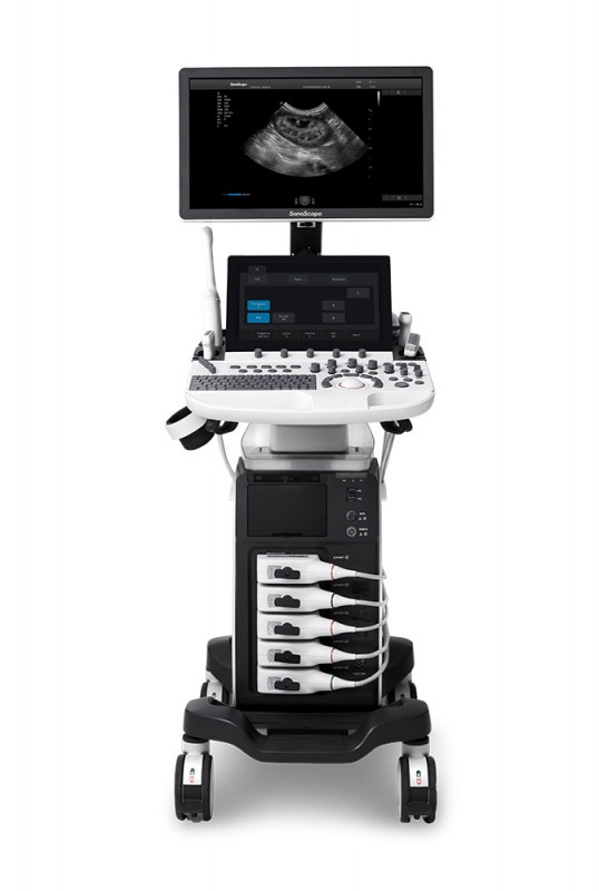
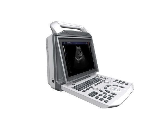
Medical ultrasound includes diagnostic techniques (mainly imaging techniques) using ultrasound, as well as therapeutic applications of ultrasound. In diagnosis, it is used to create an image of internal body structures such as tendons, muscles, joints, blood vessels, and internal organs, to measure some characteristics (e.g. distances and velocities) or to generate an informative audible sound. The usage of ultrasound to produce visual images for medicine is called medical ultrasonography or simply sonography, or echography. The practice of examining pregnant women using ultrasound is called obstetric ultrasonography, and was an early development of clinical ultrasonography. The machine used is called an ultrasound machine, a sonograph or an echograph. The visual image formed using this technique is called an ultrasonogram, a sonogram or an echogram.
Ultrasound of carotid artery
Ultrasound is composed of sound waves with frequencies greater than 20,000 Hz, which is by approximation the upper threshold of human hearing.[1] Ultrasonic images, also known as sonograms, are created by sending pulses of ultrasound into tissue using a probe. The ultrasound pulses echo off tissues with different reflection properties and are returned to the probe which records and displays them as an image.
An ultrasound result on fetal biometry printed on a piece of paper
A general-purpose ultrasonic transducer may be used for most imaging purposes but some situations may require the use of a specialized transducer. Most ultrasound examination is done using a transducer on the surface of the body, but improved visualization is often possible if a transducer can be placed inside the body. For this purpose, special-use transducers, including transvaginal, endorectal, and transesophageal transducers are commonly employed. At the extreme, very small transducers can be mounted on small diameter catheters and placed within blood vessels to image the walls and disease of those vessels.
.png)